Non ci sono recensioni
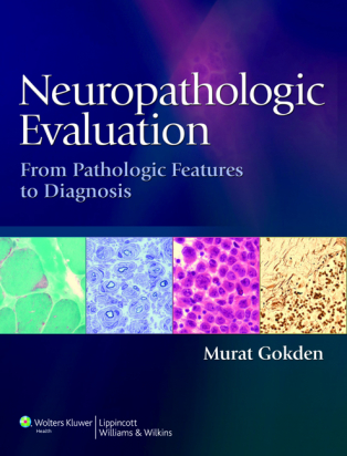
Neuropathologic Evaluation: From Pathologic Features to Diagnosis
Thorough evaluation of patient complaints, accurate interpretation of clinical signs, and assessment of gross and microscopic features are the keys to accurate neuropathologic diagnosis. With this quick reference, readers will sharpen their analytical skills and enhance their interpretive abilities—using an accessible and reader-friendly format that takes you from initial findings to differential diagnosis. The book’s broad-based coverage addresses the entire neuropathology areas, including skeletal muscle and nerve pathology, non-neoplastic neuropathology and pediatric neuropathology. An easy-to-follow format groups disease processes by common pathologic features, and an encyclopedic narrative style makes quick-referencing easy.
Look inside and explore…
• Broad-based coverage addresses the entire neuropathology areas, including skeletal muscle and nerve pathology, non-neoplastic neuropathology and pediatric neuropathology.
• Easy-to-follow format groups disease processes by common pathologic features.
• Encyclopedic narrative style makes quick-referencing easy.
• Practical approach is based on a comprehensive list of gross and microscopic features, helping you understand and interpret your findings.
Pick up your copy today!
 I- GROSS FEATURES 1. Introduction  2. Skin and External Findings  3. Bones  4. Epidural Space  5. Dura Mater and Subdural Space  6. Leptomeninges and Subarachnoid Space  7. Blood Vessels  8. Brain Weight  9. Surface Features 10. Cranial and Spinal Nerves 11. Cut Surfaces 12. Parenchymal Hemorrhage 13. Ventricular System 14. Choroid Plexus 15. Pineal Region 16. Sellar/Suprasellar Region 17. Surgical Specimens II- MICROSCOPIC FEATURES  18. Introduction  19. Unremarkable-Appearing Tissue  20. Hypercellularity  21. Lesion Borders  22. Cytoplasmic Features  23. Intracytoplasmic Inclusions  24. Mucin  25. Background and Neuropil Features  26. Accumulations  27. Calcifications  28. Pigments  29. White Matter and Myelin  30. Neuron Loss and Gliosis  31. Tract Degeneration  32. Vascular Changes  33. Perivascular Arrangements and Rosettes  34. Bone Marrow Derived Cells and Inflammation  35. Microorganisms  36. Hemorrhage  37. Necrosis  38. Cellular Arrangements  39. Papillary and Pseudopapillary Structures  40. Small Blue Cells  41. Nodular Arrangement 42. Biphasic Pattern and Composite Neoplasms 43. Cellular Enlargement 44. Nuclear Features 45. Intranuclear Inclusions 46. Mitotic Activity 47. Ependymal Surfaces 48. Choroid Plexus 49. Cystic Lesions 50. Leptomeningeal Infiltrates 51. Metaplastic Tissues 52. Pitfalls and Artifacts III- INTRAOPERATIVE CONSULTATIONS 53. Introduction 54. Cytologic Preparations  - Introduction  - Background Features  - Tissue Fragments  - Nuclear Features  - Biphasic Pattern  - Diffuse Cellularity  - Cytoplasmic Features  - Blood Vessels  - Accumulations and Structures  - Artifacts and Pitfalls  - Touch Preparations 55. Frozen Sections IV- Cerebrospinal Fluid Cytology 56. Introduction and Specimen Types 57. Gross Features 58. Background Features 59. Hematolymphoid Cells 60. Microorganisms 61. Large Single Cells 62. Cell Groups 63. Artifacts and Noncellular Material V- HISTOCHEMISTRY 64. Intruduction  65. Bielschowsky and Other Silver Stains  66. Myelin Stains  67. Reticulin Stain  68. Elastic Tissue Stains  69. Collagen Stains  70. Periodic Acid-Schiff Stain  71. Mucin Stains  72. Stains for Microorganisms  73. Stains for Pigments  74. Others VI- IMMUNOHISTOCHEMISTRY AND RELATED TECHNIQUES  75. Introduction  76. Glial Fibrillary Acidic Protein and Glial Markers  77. S-100 Protein and Melanocytic Markers  78. Neuronal and Neuroendocrine Markers   - Synaptophysin   - Neurofilament   - Neu-N   - Chromogranin   - Others  79. Epithelial Markers   - Cytokeratins   - Epithelial Membrane Antigen   - Others  80. Prognostic and Proliferative Marker   - Ki-67   - P53   - Steroid Hormone Receptors   - Others  81. Pituitary Hormones  82. Microorganism Markers  83. Hematolymphoid Markers  84. Micellaneous   - Alpha-beta-crystallin   - Amyloid   - Basement Membrane Markers   - Brachyury   - Catenin   - CD99   - Markers Used for Germ Cell Neoplasms   - INI-1   - Mesenchymal Cell Markers   - TTF-1   - WT-1   - Others  85. In-situ Hybridization  86. Immunoflueorescence Microscopy VII- RADIOLOGIC FEATURES  87. Intruduction  88. Radiologic Techniques and Terminology  89. Extra-axial Lesions  90. Dura-based Lesions  91. Meningeal Enhancement  92. Non-enhancing Lesions  93. Enhancing Lesions  94. Cystic Lesions and Mural Nodule  95. Ring Enhancement  96. Perilesional Edema  97. Leisons Crossing Midline  98. Calcification  99. Others VIII- SKELETAL MUSCLE BIOPSY SPECIMENS  100. Introduction  101. Apparently Unremarkable Tissue  102. Fiber Size Variation and Atrophy  103. Hematolymphoid Cells  104. Accumulations and Inclusions in Myofibers  105. Vacuolations  106. Internal Architecture of Myofibers  107. Necrosis  108. Nuclear Features  109. Endomysium, Perimysium, Epimysium and Blood Vessels  110. Histochemical Stains and Reactions  111. Immunohistochemistry and Immunofluorescence Microscopy IX- PERIPHERAL NERVE BIOPSY SPECIMENS  112. Introduction  113. Axons  114. Myelin  115. Blood Vessels  116. Accumulations and Inclusions  117. Inflammation and Infections  118. Schwann Cells  119. Others  120. Special Stains and Techniques X- APPENDICES
Ultimi prodotti
